Candida albicans is a polymorphic fungus that is present as a commensal in the mucosal epithelia of the human body. Changes in the host, such as immuno-suppression and loss of normal body flora promote C. albicans to transform into a pathogenic filamentous form resulting in a wide range of infection outcome- from mild, epithelial yeast infection to life-threatening infections of the heart, lungs, and blood. Gaining a deeper understanding of the biology behind the filamentation of C. albicans as well as immune response to the fungus will improve our understanding and treatment of these infections.
We model the human epithelial micro-environment by adding a solid bio-polymer substrate to C. albicans culture and controlling key environmental factors, viz., oxygen content, pH, humidity as well as mucin proteins present in the mucosal layer that lubricate and protect all internal epithelial lining, including oral, respiratory, gastro-intestinal and vaginal environments. Using different bio-polymer gel composites, we provide either a two-dimensional substrate for the cells to adhere to, or a three-dimensional porous scaffold which permits cells to move through. We can tune the mechanical properties like stiffness, and porosity of these microgel composites by tuning polymer concentrations of constituent gels to mimic different epithelia (oral, gut, etc.). We have done preliminary characterization of these composite gels, e.g., collagen+alginate and collagen+agarose, in terms of elasticity using rheology, gel morphology using confocal microscopy and cell viability using live-cell imaging.
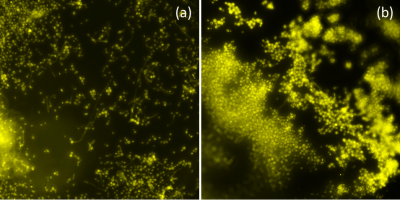
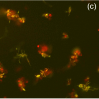
Besides providing a mechanical substrate for cell locomotion, polymer gels have other roles to play in the epithelial micro-environment. For example, the chemical composition of the constituent polymers can lead to the phenotypic changes in C albicans. We see such a phenotype suppression in C. albicans where the pathogenic filamentation is significantly suppressed in the presence of agarose gel and collagen-agarose composite. The figure above shows images of YFP-expressing C. albicans growing in well-plates, in (a) 1x PBS, and (b) embedded in a composite gel scaffold, where both samples were incubated for 6 hrs in pH 7.4 and growth medium. We see significantly less filamentation of C. albicans in the gel. With the addition of macrophages to the C. albicans however, the beneficial effect of the gel in suppressing the pathogenic phenotype is lost (above, (c)).
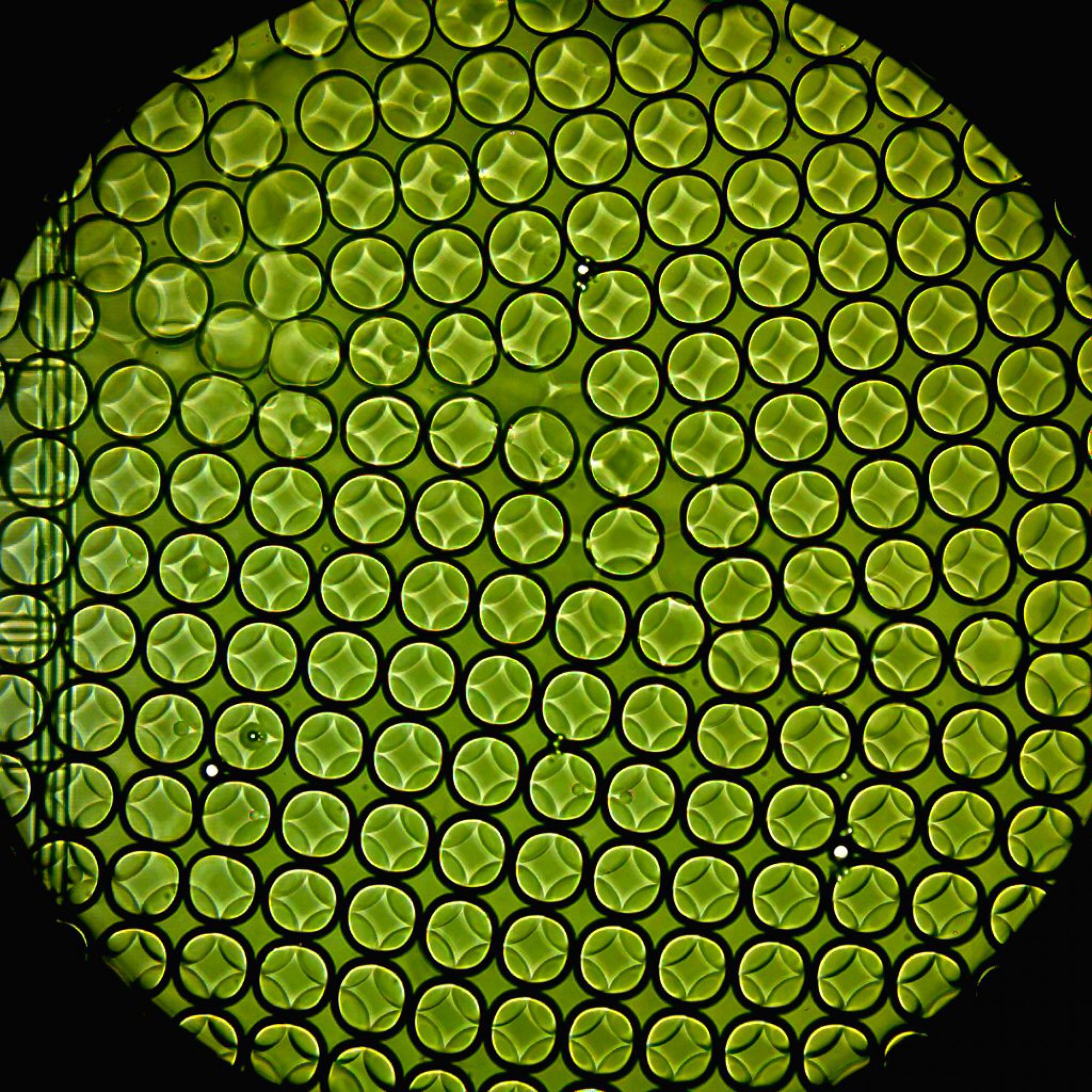
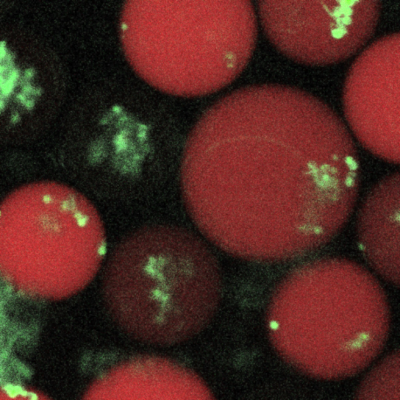 We repeat the experiment where YFP-expressing C. albicans in the yeast form is encapsulated in microgel-scaffolds in drops at different polymer concentrations (see left). We add a fluorescent dye, the concentration of which is commensurate with the gel concentration. The drops are then incubated overnight and imaged using multi-channel confocal microscopy. As before, we see a similar suppression in C. albicans growth while exhibiting the pseudo-hyphal phenotype.