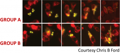
We study controlled infections of murine macrophages with Candida albicans, a medically important opportunistic fungal pathogen using reverse emulsion microfluidics. Preliminary work with this system suggests infection results in two distinct outcomes: in one, phagosome maturation successfully checks growth and results in cell death of C. albicans (GROUP A); in the other, C. albicans successfully filaments and grows within the macrophage phagosome (GROUP B), eventually killing the macrophage.
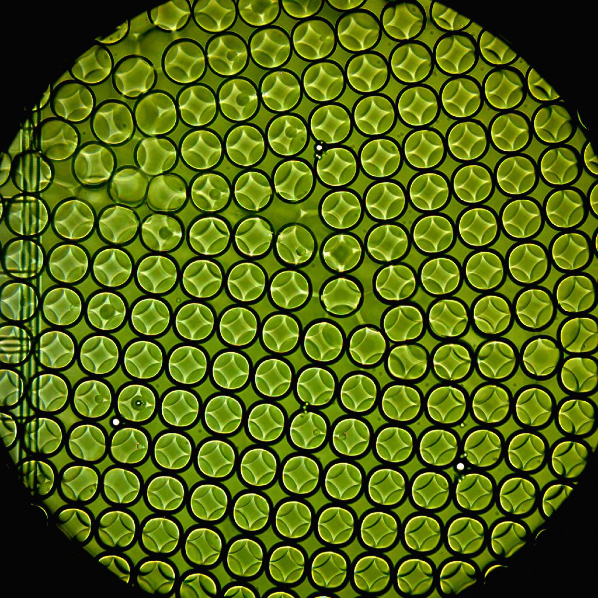
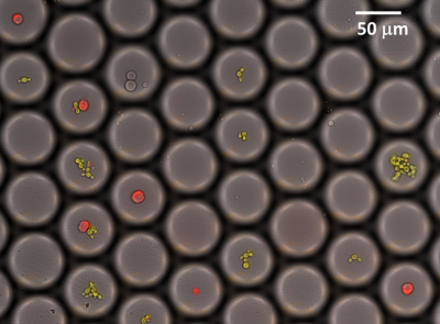
Using emulsion microfluidics to create unique, isolated micro-environment (see left), we track C. albicans and macrophage interaction at a pre-defined MOI. We then simultaneously incubate and image the host-pathogen cells in the droplets as the infection progresses, using a live-cell imaging system.
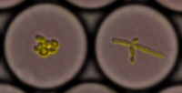
We note some interesting results: there is an intrinsic heterogeneity in the C. albicans population (yellow), as seen in the benign pseudo-hyphal cell (a) as well as the more virulent hyphal phenotype (b).
 (c) (d) (e) (f)
There are also important differences seen in the infection outcome, viz., successful phagocytosis of C. albicans (yellow) by macrophage (red) (c), different amounts of pseudo-hyphal growth in C. albicans (d & e); hyphal filaments from the phagosome growing out the macrophage, resulting in macrophage death (f).
Based on fluorescence and morphologic signal, we will sort subpopulations based on outcome using microfluidic droplet sorter. Our final aim is to perform population-level and single-cell RNA-Seq on sorted macrophages and engulfed C. albicans to transcriptionally profile cell sub-populations based on outcome- of more efficient macrophage killers (GROUP A) as well as C. albicans of greater virulence (GROUP B).


